
Ankle Sprains
Ankle sprains are the most common musculoskeletal injury, accounting for 2 million emergency department visits annually in the United States alone. Learn the anatomy, grading, and evidence-based treatment approaches for every type of ankle sprain.
Ankle Ligament Anatomy
The ankle joint is stabilized by three ligament complexes. Understanding which ligaments are involved determines the sprain type, severity, and optimal treatment approach.
Lateral Ligaments
The most commonly injured group, responsible for 85% of all ankle sprains. Injured during inversion (rolling inward).
- ATFL (Anterior Talofibular) - most commonly torn
- CFL (Calcaneofibular) - involved in Grade II+
- PTFL (Posterior Talofibular) - rarely torn alone
Medial (Deltoid) Ligament
A strong, fan-shaped ligament that resists eversion (rolling outward). Medial sprains are less common but more significant when they occur.
- Superficial and deep layers
- Injured by forced eversion or external rotation
- Often associated with fibula fractures (Maisonneuve)
Syndesmosis (High Ankle)
Connects the tibia and fibula above the ankle. High ankle sprains take significantly longer to heal than lateral sprains.
- AITFL (Anterior Inferior Tibiofibular)
- PITFL (Posterior Inferior Tibiofibular)
- Injured by dorsiflexion + external rotation
Sprain Grading System
Stretch (Mild)
Ligament fibers stretched, not tornMild tenderness and swelling. Able to bear weight. No mechanical instability detected on exam.
Partial Tear (Moderate)
Incomplete ligament ruptureModerate swelling and bruising. Difficulty with weight bearing. Mild to moderate instability on anterior drawer test.
Complete Rupture (Severe)
Full ligament tearSignificant swelling, bruising, and deformity. Unable to bear weight. Gross instability with positive anterior drawer and talar tilt.
Lateral vs. Medial vs. High Ankle Sprain
Lateral Sprain
85% of ankle sprainsMedial Sprain
5 - 10% of ankle sprainsHigh Ankle Sprain
5 - 10% of ankle sprainsOttawa Ankle Rules
The Ottawa Ankle Rules are a validated clinical decision tool used to determine when an X-ray is needed after an ankle injury. They have a sensitivity near 100% for detecting fractures, meaning a negative result reliably rules out a fracture.
- 1 Bone tenderness along the posterior edge or tip of the lateral malleolus (distal 6 cm of the fibula)
- 2 Bone tenderness along the posterior edge or tip of the medial malleolus (distal 6 cm of the tibia)
- 3 Bone tenderness at the base of the 5th metatarsal (mid-foot, outer side)
- 4 Bone tenderness at the navicular bone (mid-foot, inner side)
- 5 Inability to bear weight for 4 steps both immediately after injury and at the time of evaluation
Treatment by Grade
Functional Rehabilitation
RICE protocol for the first 48-72 hours (Rest, Ice, Compression, Elevation). Early weight bearing as tolerated with an elastic bandage or lace-up brace. Begin range-of-motion exercises within 48 hours. Progress to balance training and strengthening by day 5-7. Most athletes return to activity within 1-3 weeks.
Protected Mobilization
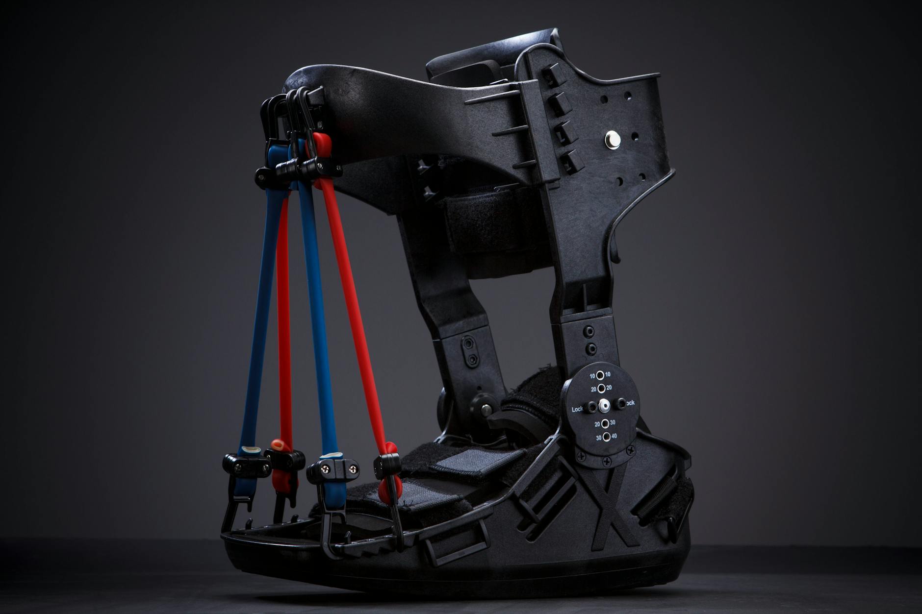
RICE protocol plus a semi-rigid ankle brace or walking boot for 2-3 weeks. Partial weight bearing initially, progressing to full as tolerated. Formal physical therapy for proprioception, peroneal strengthening, and sport-specific agility. Return to sport in 3-6 weeks with ankle bracing during activity for 6 months.
Immobilization and Possible Surgery
Short period of immobilization (walking boot or cast for 1-2 weeks) followed by protected mobilization. Surgical repair is considered for high-level athletes or patients with persistent mechanical instability after 3-6 months of rehabilitation. Comprehensive PT program lasting 8-12+ weeks. Return to sport is individualized, typically 2-4 months.
Chronic Ankle Instability
Up to 40% of people who sprain their ankle develop chronic ankle instability (CAI). This occurs when the ligaments do not heal properly or when proprioceptive deficits persist after the initial injury. Recognizing the signs early allows for targeted rehabilitation.
Repeated Sprains
Recurrent "giving way" episodes, especially on uneven terrain or during sports
Persistent Swelling
Low-grade swelling that returns after activity, even months after the initial injury
Ongoing Pain
Aching pain during or after activity, particularly with cutting, pivoting, or lateral movement
Decreased Confidence
Fear of re-injury leading to altered movement patterns and avoidance of certain activities
Proprioception Training Program
1-2
Double-Leg Balance
Stand on both feet with eyes open on a firm surface, then a foam pad. Progress to eyes closed. Hold 30 seconds, 3 sets, 2-3 times daily.
2-3
Single-Leg Stance
Single-leg balance on firm surface, then foam pad. Add eyes-closed progression. Hold 30 seconds, 3 sets each leg. Introduce ball catches for distraction.
3-4
Dynamic Balance
Single-leg balance with perturbations (partner pushes, reaching tasks). Star excursion balance test. Wobble board training, 2-3 minutes per set.
4-6
Sport-Specific Drills
Lateral hops, figure-8 runs, cutting drills on grass then court. Single-leg hop-and-hold landings. Progress to reactive agility with directional cues.
Taping vs. Bracing
Athletic Taping
- Customizable support pattern for individual injury
- Better proprioceptive feedback (skin contact)
- Lower profile inside footwear
- Can be combined with pre-wrap for comfort
- Loses 40-50% of support within 20 minutes of activity
- Requires trained athletic trainer to apply
- Higher ongoing cost (tape is single-use)
- Risk of skin irritation with repeated use
Semi-Rigid Ankle Brace
- Maintains consistent support throughout activity
- Self-applied without professional assistance
- Reusable and cost-effective long-term
- Evidence-based reduction in re-sprain rates
- Slightly bulkier inside footwear
- May restrict some dorsiflexion range
- One-size approach may not suit all injury patterns
- Some athletes report decreased agility perception